Перекрёстные ссылки книги для 1266
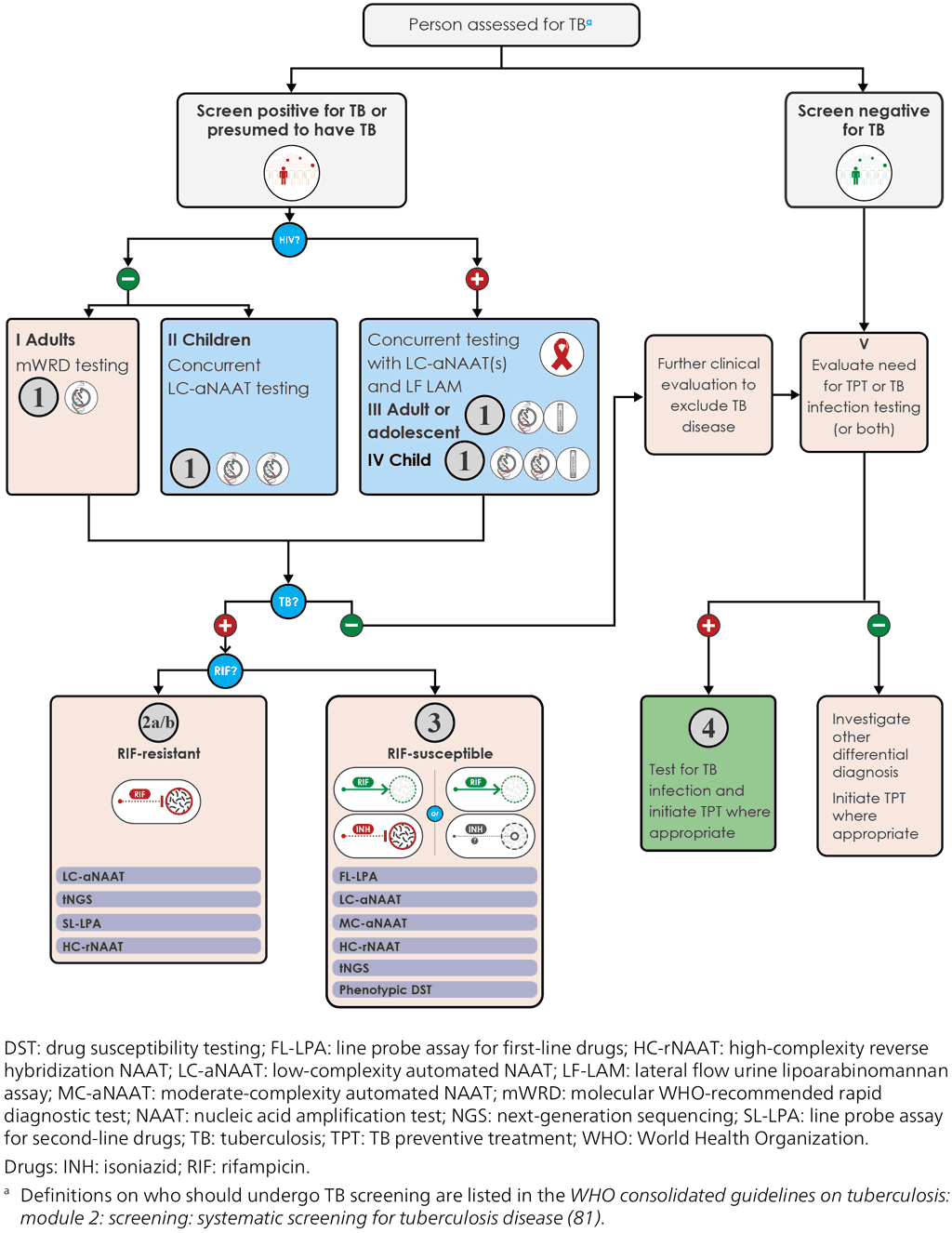
Although the algorithms are presented separately, they are interlinked and cascade from one to another. This is illustrated in the overview in Fig. 6.1, which describes how the different algorithms cascade from the starting point of a person being evaluated for TB. If the person screens positive, or is presumed to have TB, samples are collected for diagnostic and RIF-resistance testing according to Algorithm 1. The number and type of recommended samples and tests varies according to the person’s age and HIV status (as shown in Fig. 6.1: I-IV). The result of the initial test or tests will guide further actions (Fig. 6.1).
For people who test positive for TB and in whom RIF resistance is detected, further work-up is needed. Algorithms 2a and 2b provide guidance on the additional testing required to detect resistance to FQs and other second-line drugs. People with TB that is susceptible to RIF may require further testing for INH resistance and second-line drugs; Algorithm 3 provides guidance on suitable tests.
People who screen negative for TB or in whom TB disease has been ruled out through testing should be clinically evaluated. Clinical evaluation is especially important for children, who experience lower rates of bacteriological confirmation of TB owing to low and variable amounts of mycobacteria in their samples. Further practical guidance on the clinical management of childhood TB and example treatment decision algorithms can be found in the WHO operational handbook on tuberculosis: module 5: management of TB in children and adolescents (58).
People in whom TB is excluded are then assessed for the need for TPT or testing for TB infection (or both) (Fig. 6.1: V). Algorithm 4 shows the steps needed to perform testing for TB infection. As explained in Section 2.5.1, identifying, testing, evaluating and treating people with TB infection is a multistep process that has been termed the TB infection cascade of care (40). Those that do not need TPT or TB infection testing undergo evaluations for differential diagnoses.
Fig. 6.1. The cascade of the four diagnostic algorithms


 Обратная связь